The LuViva advantages
IMMEDIATE RESULTS
Results are available right at the site of examination, so prompt actions can be taken
EARLY DETECTION
May identify changes of cellular tissue than other screening methods up to 2 years earlier
HIGHER SENSITVITY
Reduces False-Negative cases missed by other procedures
EFFICIENT TREATMENT
Reduces the need of unnecessary diagnosing tests and the costs and hussles related
NON-INVASIVE METHOD
No tissue samples are required and nothing is applied to the cervix, resulting for the patient no discomfort and risk of complications
ENHANCED PATIENT CARE
Immediate emotional relief for patients
How LuViva works?
The LuViva® Advanced Cervical Scan (LuViva) works by combining fluorescence and reflectance spectroscopy to identify chemical and physical changes in uterine cervical cellular tissue. With the aid of the single-use LuViva® Cervical Guide (CG), the device painlessly scans the cervix with light for a little over a minute, analyzing the spectral data. After the scan, the results will be displayed on the monitor for immediate review by the healthcare provider. To conduct a scan and receive results, the healthcare provider is not required to apply or remove anything to the cervical tissue. The patient is simply positioned for a gynecological examination, her cervix is gently cleared of any mucous, and then a speculum and a CG are inserted. Following the prompts on the LuViva’s touchscreen monitor the healthcare provider proceeds through the test steps until the completion of the scan when the CG is removed. The LuViva’s results indicate the likelihood of cervical intraepithelial neoplasia II or greater (CIN II+) within the scanned area as Low, Moderate, or High.
Publications
For more information regarding the clinical performance of LuViva® Advanced Cervical Scan, please visit this page:
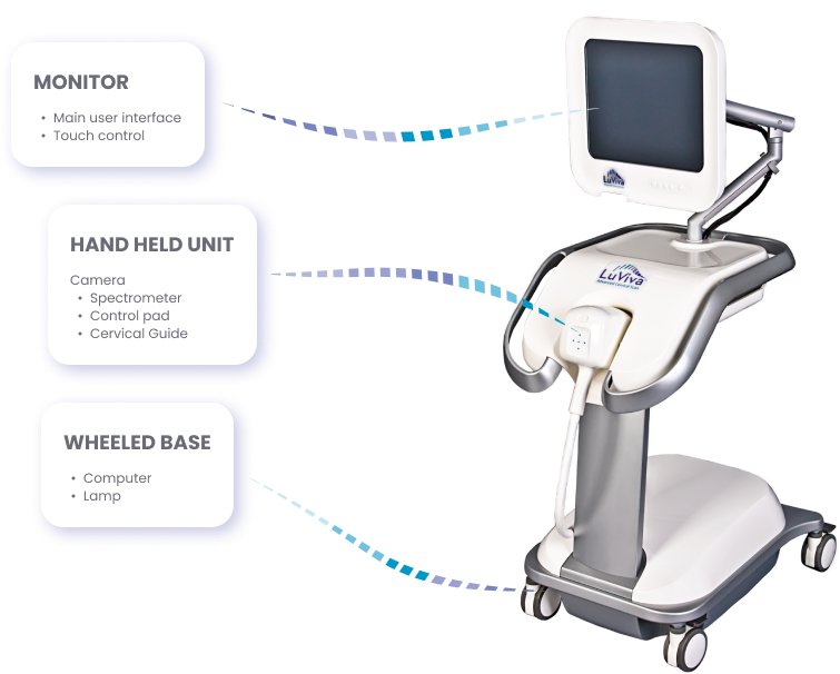
Components
The device itself consists of three main components

Certificates

FAQ
1. How is LuViva different from a Pap test?
Using light, LuViva scans the majority of cervix’s surface and penetrates 5mm below the surface, analyzing in real time the physical and chemical properties of the tissue with its spectrometer to determine the likelihood of cervical precancer or cancer.
A pap test is a cervical cancer screening test in which cells collected from the surface of the cervix are analyzed in a laboratory to determine if precancerous or cancerous cells are present.
If a lesion is not at the surface or if cells failed to be collected, disease can be missed. LuViva scans the majority of the cervix, and its light can penetrate 5 mm below the surface, so there is less of chance an important lesion will be missed.
2. How is LuViva different from an HPV test?
LuViva scans the entire cervix with light and then using its spectrometer, analyses the information in real time to determine if the physical and chemical properties indicative of cervical precancer or cancer are present.
An HPV test determines the presence of an active HPV infection in cervical tissue. While HPV is an oncovirus that is responsible for over 95% of cervical cancers, a positive HPV test does not mean cervical cancer is present.
3. What type of light is used with the LuViva and can it harm the patient or healthcare workers?
LuViva is compliant with all U.S. and international guidelines regarding exposure to light and is similar to the amount of ultraviolet or other light generated by a typical colposcopic examination. LuViva uses UV/visible light from 300 to 800 nm and it is delivered to the patient and only the patient via the HHU through the Cervical Guide.
4. Does the CG come in any other sizes or colors?
No. The CG comes in only one size and color. In order to accommodate the majority of women and allow for enough of the transformation zone to be scanned, the width of the CG is 2.5cm, which is the average width of women’s cervixes. The LuViva’s camera is preset to focus at the distance from the HHU to the CG’s distal end. Additionally the spectrograph is programmed to take measurements from this distance. The CG’s color is black to block light and prevent any spectral interference when scanning.
5. Can LuViva detect any other diseases or cancers?
No. The LuViva is designed only to identify the physical and chemical indications of cervical cancer.
6. Is the CG reusable?
No, for patient safety and optimum test results the Cervical Guide is not reusable.
7. Will the LuViva identify where a physician should take a biopsy?
No, LuViva only indicates the likelihood that CIN2+ is developing within the cervical tissue.
8. Does the device scan the entire cervix or only certain areas?
LuViva scans the entire area within the 2.5 cm opening of the CG centered on the os.
9. Does LuViva scan the cells of the ectocervix, endocervix, or both?
The LuViva scans both the ectocervix and part of the endocervix closest to the ectocervix. Epithelial tissue models indicate that the light used by LuViva penetrates approximately 5 mm into the cervical epithelial layer. This also applies to how far the light travels into the endocervix. In clinical trials, LuViva was shown to successfully detect CIN2+ in the endocervix. In our US study (Twiggs et al, 2013), there were 23 such cases. This supports the tissue models that indicate that LuViva can scan both the ectocervix and at a minimum, the distal endocervix.
10. How do I prepare the patient for a LuViva scan?
Once the speculum is inserted, gently remove any mucous and blood from the cervix with a gauze or large cotton swab. Do NOT apply anything to the cervix. Do not apply lubricant to the CG.
11. Can a Pap/HPV/VIA/VILLA test be performed on the same day as a LuViva scan?
Due to physical disruptions and, if applicable, the application of acetic acid or iodine, Pap/HPV/VIA/VILLA tests should only be done AFTER the LuViva scan.